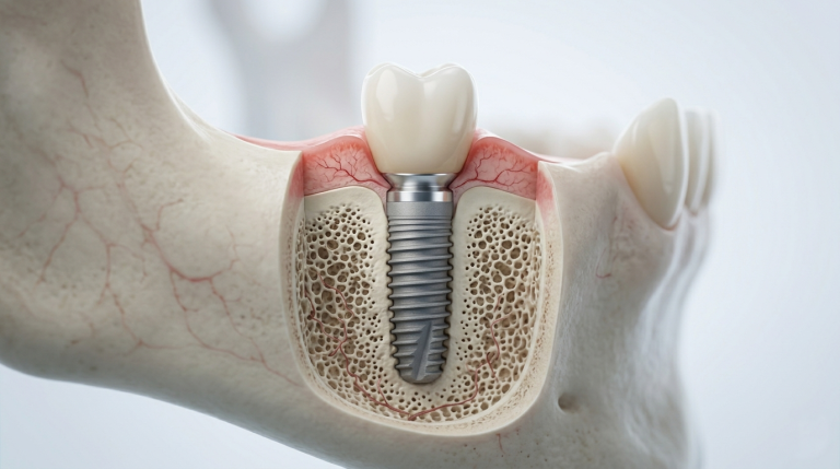
Dental Implants vs. Dentures: Which Is the Better Long-Term Choice?
Missing teeth can affect more than your smile; they can impact your confidence, speech, eating habits, and long-term oral health. This guide...

Flat X-rays show a two-dimensional image of a three-dimensional structure. For complex procedures like dental implant placement, wisdom tooth removal, or airway evaluation, that limitation matters. Cone beam 3D imaging, or CBCT, gives Dr. Joshua Englander a precise, three-dimensional view of your teeth, roots, jawbone, nerves, and sinuses in a single scan so every treatment decision is based on complete information.

Modern Tech
Precise, efficient care
Cone beam computed tomography is a specialized X-ray system that rotates around the patient and captures hundreds of images from different angles, which are then reconstructed into a detailed 3D volume by computer software. Unlike a standard medical CT scanner, the cone beam system uses a focused, cone-shaped beam that limits radiation exposure to the area of interest. The result is a highly detailed 3D model that shows bone density, root anatomy, nerve pathways, sinus cavities, and the precise relationship between teeth and surrounding structures, none of which are fully visible on a flat X-ray.
Dr. Englander uses cone beam imaging for cases where two-dimensional imaging is not sufficient to plan treatment safely and accurately. The most common applications include dental implant planning, where the scan confirms exact bone volume and the location of the inferior alveolar nerve before an implant post is placed. It is also used to evaluate wisdom teeth and their proximity to the nerve canal, to diagnose TMJ joint conditions, to assess airway anatomy, to plan bone grafting procedures, and to investigate the source of pain or pathology that is not visible on standard X-rays.
For patients considering dental implants, cone beam imaging is transformative. The scan allows Dr. Englander to measure bone depth and width at the exact proposed implant site, identify the precise position of nerves and blood vessels, detect bone deficiencies that would require grafting before implant placement, design a surgical guide that directs the implant drill to exactly the right angle and depth, and predict the final aesthetic outcome before any surgery begins. Patients who receive implants planned with 3D imaging have significantly lower rates of complications and more predictable healing.
Yes. The radiation dose from a cone beam scan is a fraction of a conventional medical CT scan. Dr. Englander follows the ALARA principle, meaning imaging is ordered only when the clinical benefit clearly justifies the minimal exposure. For most routine checkup patients, standard digital X-rays are sufficient. Cone beam imaging is reserved for complex cases where the 3D information changes the treatment plan in a meaningful way.

We Welcome You to Elevated Smiles Dentistry of Bel Air Maryland.
Schedule an AppointmentVisit us at our modern office in downtown Bel Air. We’re ready to help you achieve your best smile.
Monday: 8am – 4pm
Tuesday: 8am – 4pm
Wednesday: 7am – 3pm
Thursday: 7am – 3pm
Friday: By Appointment Only
Saturday: Closed
Sunday: Closed
Stay informed with the latest dental health insights and tips from our team.
Missing teeth can affect more than your smile; they can impact your confidence, speech, eating habits, and long-term oral health. This guide...

Veneers vs. crowns — learn the differences in coverage, durability, and cost so you can choose the right restoration for your smile...

Wondering if Invisalign can fix your overbite? Learn how clear aligners correct mild to moderate overbites and when traditional braces or surgery...

Veneers can dramatically improve the appearance of your smile, but many patients wonder how long they actually last. This guide explains veneer...

Learn how long Invisalign takes for adults and teens, what affects treatment time, and what to expect during your smile journey in...
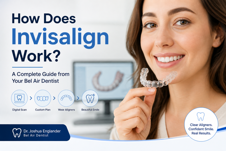
Learn how Invisalign works, what to expect with pain, and treatment steps from a Bel Air dentist. Simple, clear answers to help...